UvA researchers build brain atlas: “We still understand little of what psychological symptoms look like in the brain”

A psychosis or depression can currently only be diagnosed based on symptoms. What changes in the brain is still unknown territory. UvA neurobiologists want to change that with a brain atlas at the cellular level.
“Which brain would you rather have?” Inge Huitinga, Associate Professor of Neuroimmunology at UvA's Swammerdam Institute, looks at a pair of brains in alcohol. One brain is smaller and has much thinner coils than the other.
“The shriveled brain belongs to an Alzheimer’s patient,” Huitinga explains. “We now know quite a bit about what goes wrong in the brain with that disease. This brain, on the other hand,” she says, pointing to the healthy one, “could just as well be the brain of a psychiatric patient. You can’t see any of that with the naked eye.”
That is one reason why brain research into psychiatric syndromes such as psychosis, schizophrenia, and depression has lagged behind neurological diseases like Alzheimer's, MS, and Parkinson’s. Huitinga explains: “We know absolutely nothing about the cellular and molecular changes that lead to depression or psychosis. Absolutely zero.”
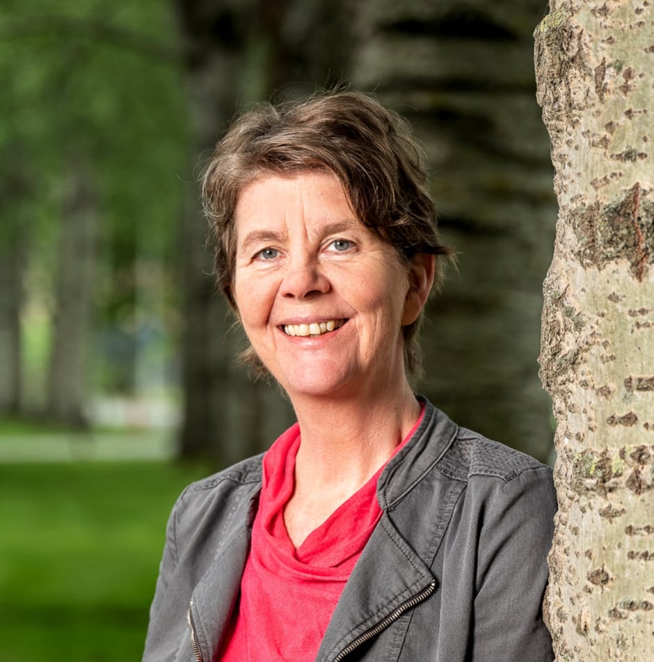
To catch up, neurobiologists, psychiatrists, chemists, and computer scientists have joined forces in the Institute for Chemical Neuroscience (iCNS). At the end of March, the consortium, led by Inge Huitinga (UvA/NIN), Paul Lucassen (UvA), and Mario van der Stelt (RUL), received a €23 million research grant to find answers to the question of how cells in the brain change in conjunction with psychiatric symptoms such as anxiety and depression.
Psychiatric brain bank
“The starting point is the human brain,” Huitinga says in her office at the Netherlands Brain Institute (NIN-KNAW) in Amsterdam. “Most researchers use questionnaires, brain scans, or laboratory animal models to investigate symptoms of depression or anxiety, and later test the results on the human brain. The problem with that is that you can make a mouse afraid, but not suicidal. We want to do it the other way around. We start in the human brain - in humans themselves (in vivo) and in the lab (in vitro) - and later validate those results in laboratory animal models.”
The Dutch Brain Bank, situated in the Netherlands Brain Institute, has received well-documented brain tissue from 5,000 donors with and without neurological syndromes since its inception in 1985. Through a special donor program for psychiatric syndromes that started in 2013, well-documented brain tissue from people with such syndromes is now also available for research.
With this, iCNS researchers can now get to work. They are not looking at it on a diagnosis-by-diagnosis basis but on a symptom-by-symptom basis. A diagnosis is made by a psychiatrist based on the patient's accounts. That results in very heterogeneous patient groups, so comparing that group with healthy people (control groups) yields little result.
With the help of AI, the researchers were able to distill individual symptoms such as anxiety and depression from the donors’ documentation. Huitinga explains: “We know which areas in the brain control the function that becomes disturbed with psychiatric symptoms. So we can we can start looking in those areas at what has changed for each symptom.”
All in your head
With this research, anxiety disorders and depression could possibly be physically demonstrated in the future, thus alleviating the stigma associated with mental illness, Huitinga hopes. “When someone is anxious, people sometimes say, ‘It's all in your head.’ But that's literally where it is. When you're anxious, your nerve cells connect differently in certain areas. That's why I like to show the brain to people to demonstrate where the symptoms come from.”
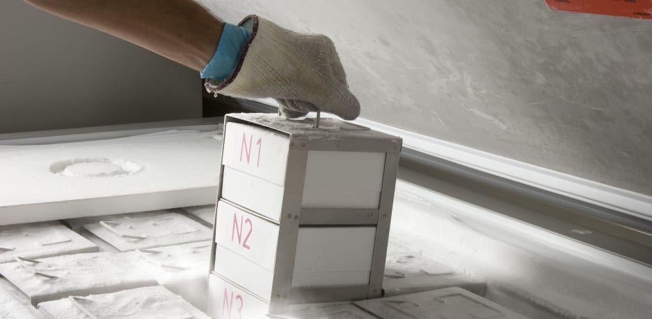
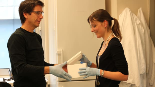
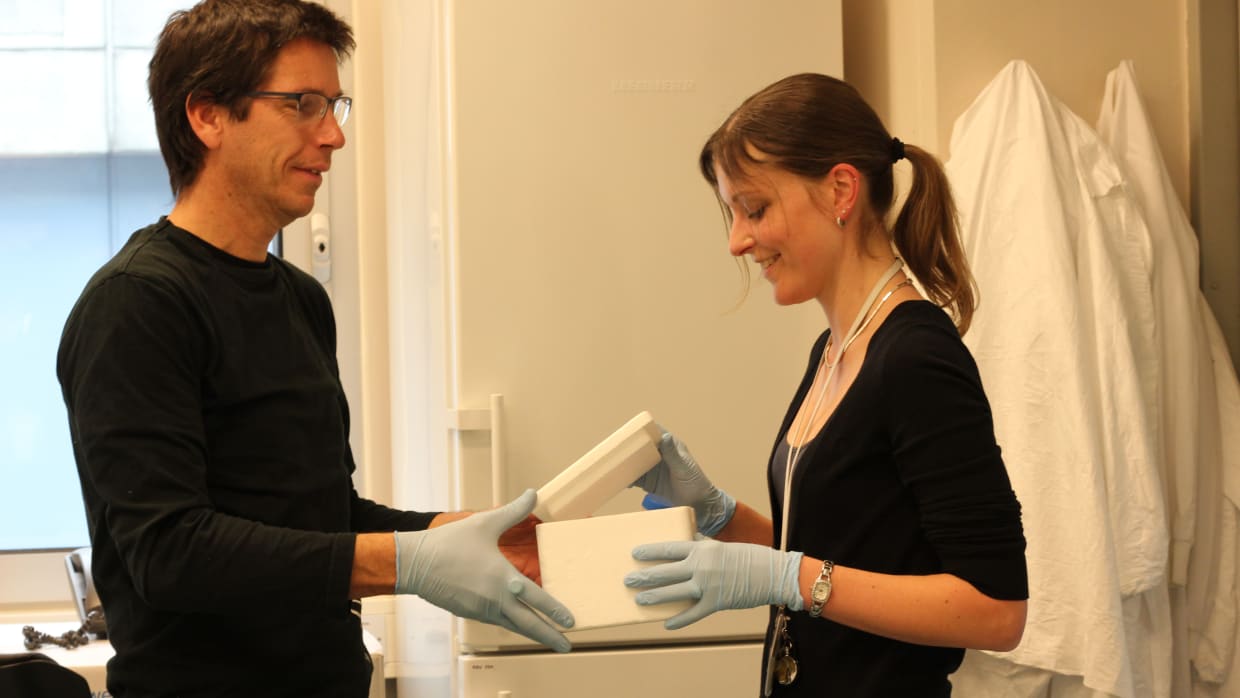
Huitinga walks from her office into the deep freeze, which has a dozen giant freezers kept permanently at -70 degrees Celsius. She removes a frozen pink brain in a vacuum bag from one of the freezers. “This is how it looks while it’s still recognizable. But when a donor comes to the Dutch Brain Bank, up to 200 areas are taken from the brain and frozen separately.” Huitinga takes out a square white box from the freezer containing a bunch of numbered trays. She opens a box. In it is a small salmon-colored piece of brain. “With this brain area, you can smell, for example.”
The iCNS researchers study 16 areas of the brain that are relevant to psychological symptoms. “Anxiety is regulated by the amygdala, the almond nucleus. So when someone has an anxiety disorder you start looking in that area.”
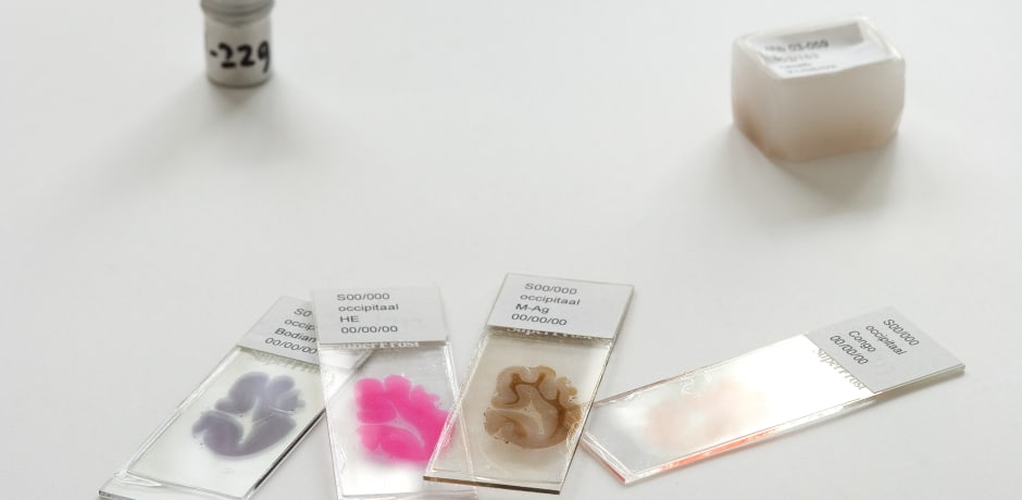
To do this, the brain tissue is first fixed in a kind of candle wax from which very thin slices of 1/6000th of a millimeter are cut. With these slices and using many new chemical techniques, researchers can see under the microscope which molecules, proteins, and fats are present in each cell. “Everything is coming together now. The data is there, the chemistry is ready, so we can now cut loose.”
Brain atlas
Back in her study, Huitinga demonstrates what that looks like. On her computer screen are the colored brain islets, enlargements of the slices under the microscope. She types in the name of a molecule on her computer screen and presses enter. Green dots light up all over the screen. “That's what I mean by a brain atlas. We can see where a molecule is in each cell.”
For example, when the researchers track down a particular molecule, they can start examining it in people with psychiatric illnesses in the clinic. They can detect the molecule in the blood or label it with another fluorescent molecule, making it detectable on a PET scan of the brain. Drugs could then emerge from that research, although that is still in the future. Huitinga says: “I think we can be satisfied if in 10 years we can make a proper diagnosis.”